Waiting for your mammography screening results can be anxiety-inducing, and we all hope that everything is normal. If your mammogram lay letter indicates that follow-up is needed, it’s easy to immediately assume the worst, but an abnormal mammogram might not be anything to worry about. Read on to learn what to do if your radiologist finds a lump during your mammogram.
What if a lump is found during a mammogram?
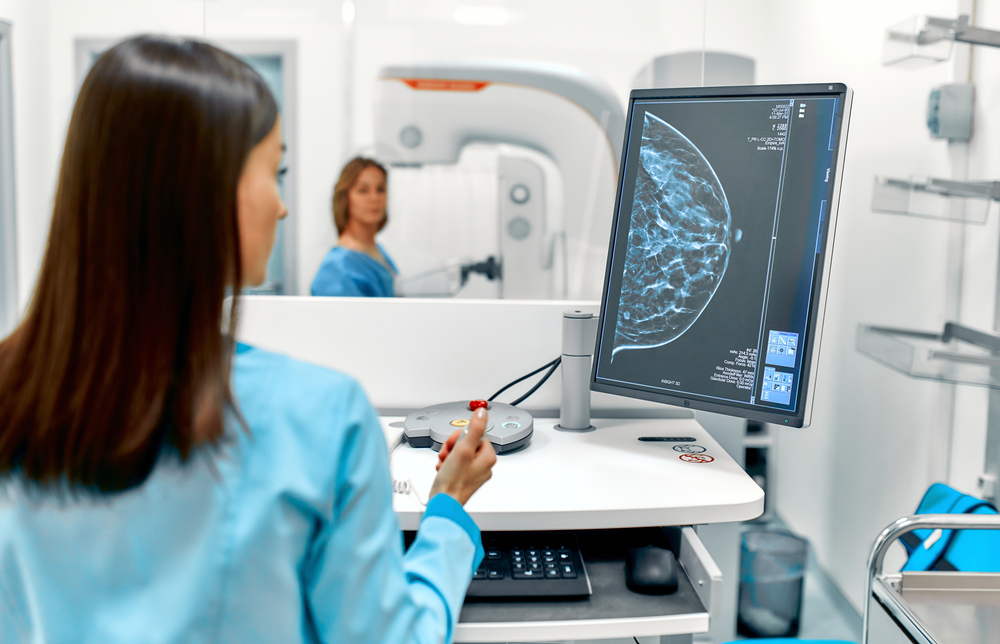
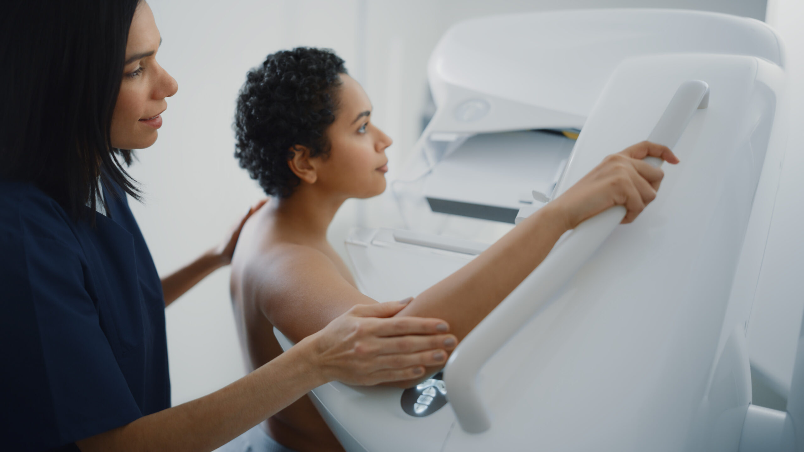
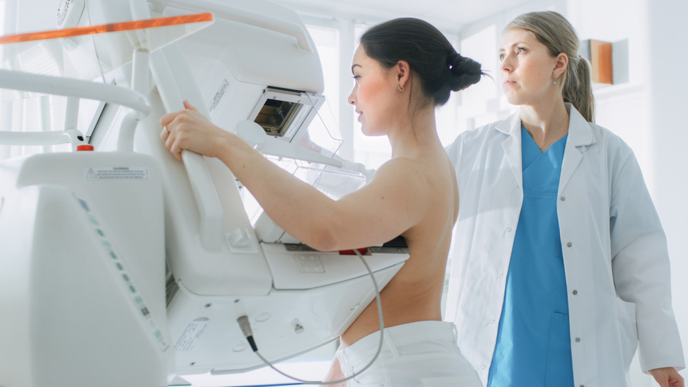
The first and most important thing to remember is that mammography screening is a detection tool, but a mammogram does not diagnose cancer. A mammogram is an x-ray picture of the breasts that allows your doctor to look for lumps and other abnormalities so that further testing can be done if necessary. The reality is that most abnormal mammograms do not end up being cancer, but early detection is your best tool for optimal health outcomes, so it’s always better to know as soon as possible.
What causes an abnormal mammogram?
There are many reasons that your mammography screening could come back as abnormal and have nothing to do with cancer. Radiologists assign each mammography screening a BI-RADS category from 0 to 6 based on the results. The most common reasons for an abnormal or inconclusive mammography screening are:
Unclear images
If the mammogram results are not clear, the doctor may want to try a second time, just to be on the safe side
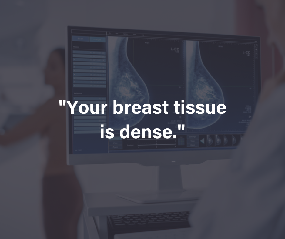
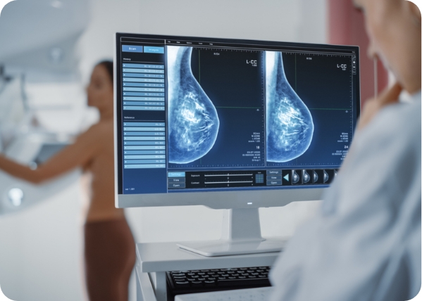
Dense breast tissue
If you have dense breasts, it may be more difficult to get a clear image, and your doctor may want you to do another mammogram or try another imaging method listed below
Non Cancerous mass or cyst:
Most breast lumps are benign (noncancerous), but it’s important to get them checked out to be sure. Common benign lumps that show up in breast imaging are:
- Fibroadenoma: A very common painless lump in breast tissue.
- Cyst: Fluid-filled sac in breast tissue that may feel soft or hard.
- Abscess: Pus pocket in breast that causes fatigue, soreness, fever, and inflammation.
- Galactocele: Fluid-filled mass usually caused by a blocked milk duct.
- Fat necrosis: Painless, hard, and firm lumps caused by damaged or disintegrating breast tissue from a bruise, lumpectomy, or radiation from previous cancer treatment.
- Hematoma: Blood-filled mass commonly resulting from surgery or injury to the breast.
- Sclerosing adenosis: This often painful excess growth of tissue in the breasts may calcify and present as lumps in a mammography screening.
Mammography Screening Follow Up Tests
If your mammogram is abnormal or inconclusive, your doctor will likely refer you for one or more of the following types of tests to confirm or eliminate a cancer diagnosis:
3-D mammogram
A 3-D mammogram is a particularly helpful type of breast imaging for women with dense breasts. This method takes 3-D images of the breast from multiple views, allowing your doctor to see the fine details more clearly.
Breast MRI
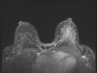
Breast MRI (magnetic resonance imaging) takes detailed pictures of the breasts using strong magnets and radio waves. Breast MRI is typically used in addition to mammograms for women with a higher risk of breast cancer, rather than being used alone.
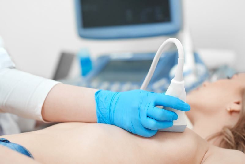
Breast ultrasound
A breast ultrasound is not typically considered to be a breast cancer screening tool. This procedure uses soundwaves rather than radiation to take a closer look at the breasts, making it a good follow-up tool for suspicious masses as well as a safer option for pregnant women.
Breast biopsy
A biopsy is the only way to confirm cancer with 100% certainty. Your doctor will use a needle to remove a very small sample of the suspicious breast tissue found with other breast imaging to be absolutely sure if cancer is present or if the mass is benign.
What if it’s cancer?
Even if the lump does turn out to be cancer, there’s not necessarily any reason to panic. Detecting breast cancer in its earliest stages makes the survival rate nearly 100%. If further testing confirms the presence of cancer, your doctor will refer you to a breast surgeon to determine the appropriate next steps to help you achieve optimal health outcomes.
Take Control Of Your Breast Health
Mammography screening can be scary, but early detection is the best way to protect your breast health, whether or not cancer is present. MagView remains committed to providing cutting-edge breast imaging technology for radiologists and to educating and empowering women on their health journey. Follow our women’s health blog for more information on breast cancer and other issues that impact your health.
Reference:
- https://www.mayoclinic.org/diseases-conditions/suspicious-breast-lumps/symptoms-causes/syc-20352786
- https://www.acr.org/Clinical-Resources/Reporting-and-Data-Systems/Bi-Rads#:~:text=BI%2DRADS%20reporting%20enables%20radiologists,assessment%20and%20specific%20management%20recommendations.&text=The%20BI%2DRADS%20atlas%20includes,includes%20mammography%2C%20ultrasound%20and%20MRI
- https://www.cancer.gov/types/breast/breast-changes
- https://www.cancer.org/cancer/types/breast-cancer/understanding-a-breast-cancer-diagnosis/breast-cancer-survival-rates.html
- https://www.komen.org/breast-cancer/screening/follow-up/










![monitoring breast density shutterstock_1299510538-[Converted]](https://magview.com/wp-content/uploads/2023/05/shutterstock_1299510538-Converted.jpg)
